This is an open access article distributed under the terms of the Creative Commons Attribution Non-Commercial License (https://creativecommons.org/licenses/by-nc/4.0/) which permits unrestricted non-commercial use, distribution, and reproduction in any medium, provided the original work is properly cited.
Adolescent idiopathic scoliosis (AIS) is the most common spinal deformity in adolescents, affecting individuals aged 10–16, with a multifactorial etiology involving genetic, hormonal, and biomechanical influences. This comprehensive literature review included 112 studies from journal conception to February 2025, retrieved from PubMed, Ovid MEDLINE, Scopus, and the Cochrane Library, focusing on epidemiology, pathophysiology, and treatment outcomes. AIS predominantly affects females, with a sex ratio increasing up to 7:1 in severe cases (>40° curves), and with right thoracic curves being the most common pattern. While bracing effectively prevents curve progression in moderate cases, spinal fusion remains the gold standard for severe deformities, ensuring long-term correction and preventing progression, despite a reduction in flexibility. Most adolescents return to full activities within a year postoperatively, with a low incidence of complications, including neurologic injury (~0.2%), infection (1%–5%), and pseudarthrosis (~1%). Future research will focus on optimizing non-fusion therapies, enhancing screening strategies, and integrating personalized medicine to improve patient outcomes and quality of life.
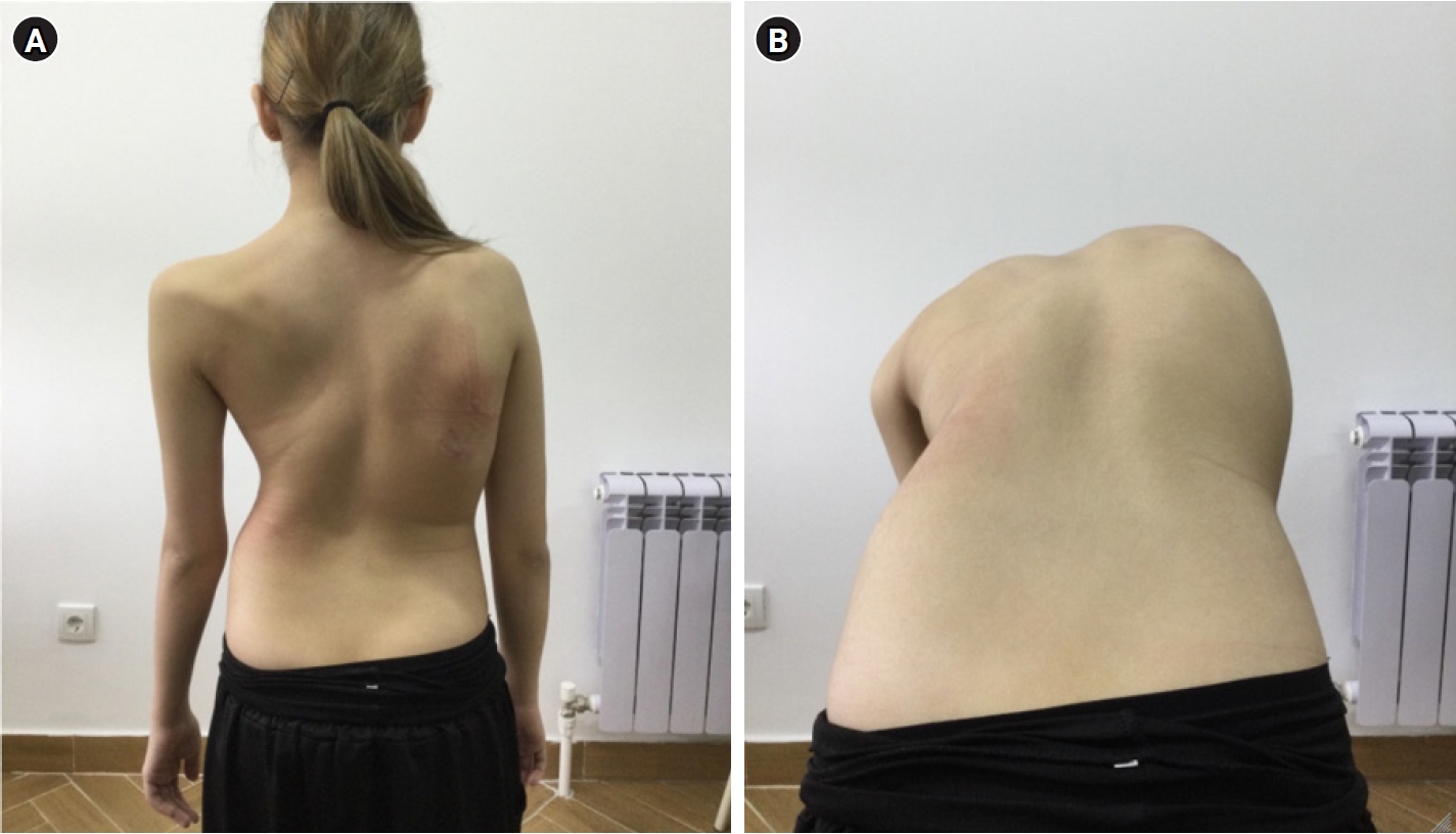
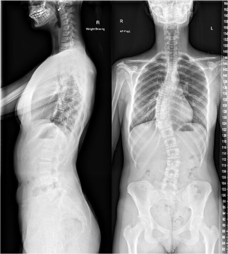
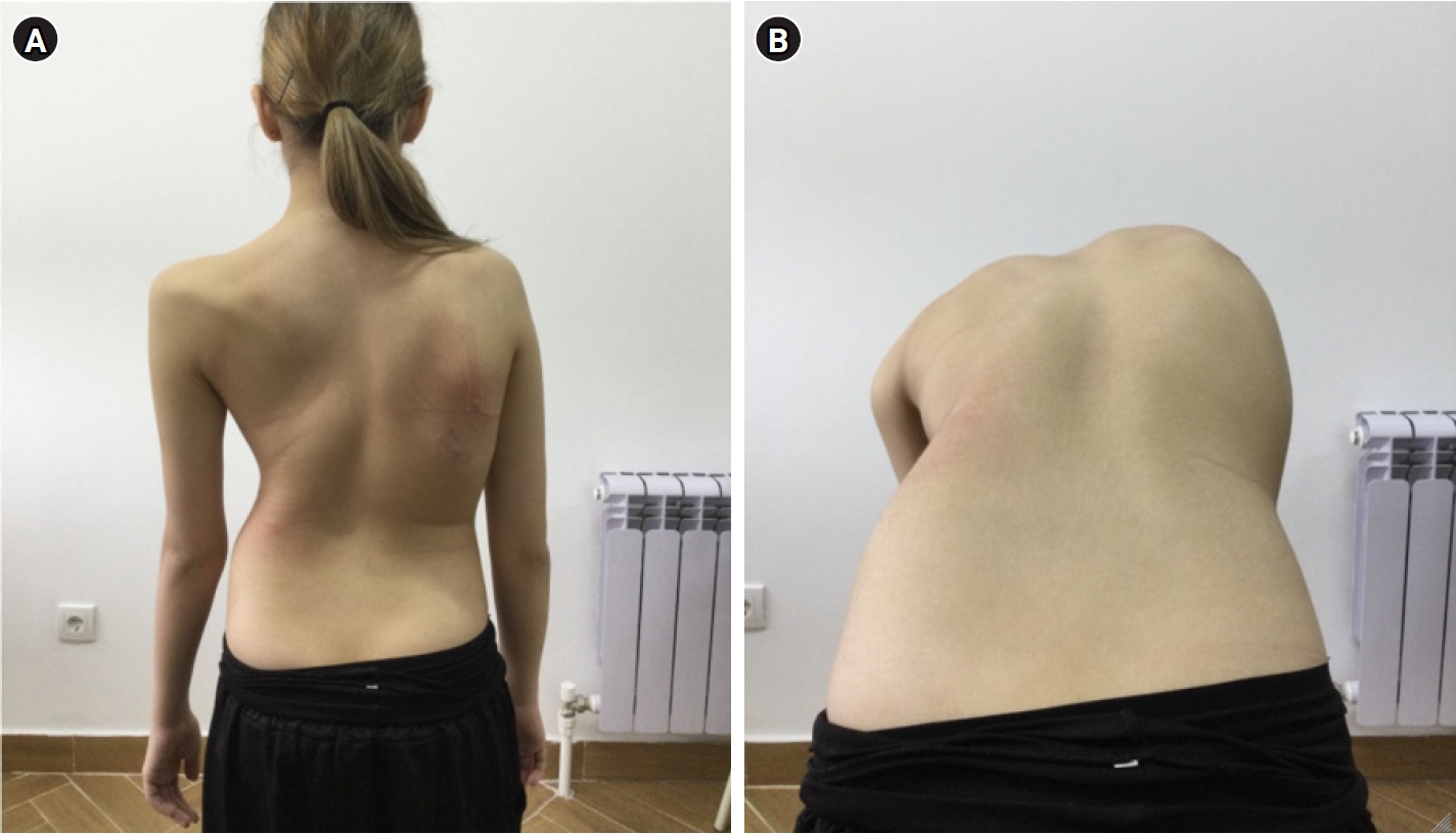
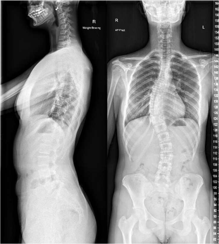
Adolescent idiopathic scoliosis (AIS) is the most common form of scoliosis, characterized by a lateral curvature of the spine ≥10° in the coronal plane with vertebral rotation in patients aged 10–16 years.1,2) It accounts for ~85%–90% of pediatric scoliosis cases.3) AIS has an estimated 2%–4% prevalence among adolescents.3) While many curves remain small and asymptomatic, a subset will progress which may lead to complications such as rib cage deformity, pulmonary compromise, chronic back pain, degenerative disc disease, and psychological distress.3-6) Typical clinical and radiographic appearances of AIS are shown in Figs. 1 and 2.
Females and males have similar prevalence of mild curves, but females have a much higher risk of progressive deformity (up to 5–10 times greater).3) This female predominance in curve progression underlies the clinical importance of early detection and monitoring in at-risk patients. Despite decades of study, the exact etiopathogenesis of AIS remains elusive, representing a major knowledge gap. AIS is deemed “idiopathic” because no definite underlying cause is identified. Current hypotheses implicate genetic factors, growth and hormonal influences, biomechanical imbalance, and environmental contributors, but no single unifying mechanism has been confirmed.1) This uncertainty makes it challenging to predict which children will develop significant curvature and when intervention is optimal. Another debated area is screening and early detection. Routine school scoliosis screening was once widespread, but guidelines diverge on its value, with various nations opting for and against nationwide screening due to conflicting evidence surrounding the fear of overdiagnosis and treatment with excessive detection, the lack of standardized testing and the cost-effectiveness of strategies.7)
AIS can have profound long-term impacts if progressive. Curves exceeding ~50° are more likely to worsen even after skeletal maturity, potentially causing pain and restrictive lung disease in adulthood.8-10) Even moderate deformities can affect adolescents’ body image and quality of life; postural abnormalities have a profound effect on body image at an important stage in psychological development, with an inverse correlation between Cobb angle and self-image.11)
Early identification and management of AIS can prevent severe deformity and the need for extensive surgery. However, determining the optimal timing and modality of treatment (observation vs. bracing vs. surgery) is a clinical challenge. Overtreatment of mild curves can subject patients to unnecessary bracing, whereas undertreatment of progressive curves may miss the window for less invasive correction.3)
Methods
A comprehensive literature review was conducted to gather evidence on AIS epidemiology, etiology, and management from journal conception to February 2025. The primary databases searched included PubMed, Ovid MEDLINE, Scopus, and the Cochrane Library. Search terms such as “adolescent idiopathic scoliosis,” “AIS epidemiology,” “AIS genetics,” “scoliosis bracing,” and “AIS surgery outcomes” were used in various combinations. Reference lists of relevant articles were also cross-checked to identify additional studies. A total of 112 papers were included in this review. Key inclusion criteria were as follows: (1) studies focusing on adolescent patients (ages 10–18) with idiopathic scoliosis (excluding congenital or neuromuscular scoliosis), and (2) those reporting on prevalence, risk factors, pathophysiology, or outcomes of treatment (observation, bracing, or surgery). Both English-language and high-quality translated articles were included.
Epidemiology
1. Prevalence
AIS is relatively common in the pediatric population. Reported prevalence in adolescents ranges from approximately 0.5% to 5.2% worldwide.1,12) This variation depends on the population studied and the screening methods used. Large school screening programs have found prevalence around 2%–3% in many regions. For example, a cohort study in Hong Kong reported scoliosis (curve >10°) in 2.5% of 157,444 students.13) Similarly, rates of about 2%–3% have been observed in other Asian populations.14)Table 1 summarizes epidemiological data on AIS prevalence and demographics. Notably, prevalence tends to increase with age during the adolescent growth spurt; one study found the prevalence of scoliosis rose from ~1% at age 11–12 up to over 3% by age 16–17 in screened schoolchildren.15) This aligns with the timing of pubertal growth, as curves often manifest or worsen during periods of rapid spinal growth.
2. Sex differences
Mild spinal curvatures (10–20° Cobb) are found in both sexes at similar frequency.3,12) However, female patients have a significantly higher risk of curve progression and development of larger curves. Epidemiological data indicate an overall female-to-male ratio of about 1.5–2:1 for AIS prevalence, which increases with curve severity.12) For curves >30°, and especially those warranting treatment, female predominance is much more pronounced. One review found the ratio of females to males was ~7:1 for curves >40°.16) In other words, while boys and girls may develop small curves equally, girls are far more likely to develop clinically significant scoliosis. Clinically, this means adolescent girls, particularly around early puberty, represent the highest-risk group for AIS detection and monitoring.
3. Risk factors
Aside from female sex, the strongest risk factor for AIS is a positive family history of scoliosis. A significant proportion of AIS patients have a family member who also had scoliosis—one study noted up to 51% of AIS patients had one or more affected relatives.17) Twin and family studies support a heritable component: if both parents had idiopathic scoliosis, their children have an estimated 50-fold higher likelihood of requiring treatment for scoliosis.18) These data underscore the genetic predisposition in AIS. Other risk factors and associations include age and growth velocity.19) There is some evidence that low body mass index and delayed skeletal maturity correlate with scoliosis progression, possibly due to prolonged growth periods or hormonal differences.20)
Etiology
The precise cause of AIS is multifactorial and remains incompletely understood.1) By definition, AIS has no clear underlying condition causing the curvature (such as vertebral malformations, neuromuscular disease, or connective tissue disorder). Instead, research suggests a convergence of genetic predisposition and environmental or biomechanical triggers. In essence, AIS has been hypothesized to involve a delicate imbalance in the growing spine influenced by hereditary factors and modulated by growth and hormones.
1. Heritability
AIS has a well-recognized familial tendency. A large-scale genetic epidemiology study of 415 Chinese female AIS patients estimated the heritability of AIS at 87.5%.21) Monozygotic (identical) twins show much higher concordance for scoliosis than dizygotic twins, indicating that genes play a significant role.22,23) Family aggregation studies have reported various inheritance patterns, and no single Mendelian pattern fits all, which points to AIS being a polygenic disorder.24) Having an affected first-degree relative greatly increases a child’s risk of AIS.
2. Genetic loci and genes identified
In the past decade, there have been 30 genetic loci identified to be associated with AIS, although each contributes only a small effect on risk.25,26) One of the earliest and most replicated findings involves the LBX1 gene on chromosome 10q24.31, with a variant near LBX1 (rs11190870) identified as a major susceptibility locus, especially in females.27)LBX1 is involved in embryonic development of muscle and the nervous system, and its role in AIS may relate to subtle neuromuscular imbalances in the spine.27) Other consistently reported genes are listed in Table 2.27-35)
Pathophysiology
The emerging picture is that genetic factors set the stage for AIS by creating a susceptibility - for example, subtle differences in vertebral growth regulation, intervertebral disc composition, or neurosensory feedback. The exact disease process remains undefined with a range of hypotheses emerging based on a complex interplay between initiation and deformity progression.
1. Genetic testing and counseling
Given the polygenic nature, routine genetic testing for AIS is not yet part of standard care; research tests exist, but the genetic variability associated with AIS pathogenesis means it is difficult for standardized testing to be performed. As more AIS-associated variants are discovered, polygenic risk scores will likely improve, however it is not possible to accurately predict which patients will develop a significant progressive deformities. Many studies have been undertaken to develop methods of predicting disease progression, suggesting further comprehensive and multi-center studies are required to develop reliable models.36)
2. Pubertal growth
AIS typically manifests during early puberty, when rapid skeletal growth occurs. Girls often develop scoliosis just before or around their peak growth velocity and before menarche. Later age of menarche has been associated with higher prevalence of AIS37), with low estrogen levels leading to reduced bone mineralization and thus an increased risk of bone deformity38). There is a complex interplay of abnormal levels of many hormones including estrogen, melatonin, growth hormone, leptin, adiponectin and ghrelin, which likely contribute to the occurrence and development of AIS in this peak growth period.39)
3. Biomechanical factors
Asymmetric loading is widely acknowledged as a key driver of curve progression, consistent with the Huerter-Volkmann principle whereby mechanical compression inhibits, and tension promotes growth of the vertebral end plates.40,41) Increased compressive forced on the concave side of the curve mediate inhibitory modulation of endochondral vertebral and associated discal growth, while reduced loading on the convex side permits continued elongation. During periods of rapid growth this imbalance can establish a self-reinforcing cycle of spinal curvature and axial rotation.10) Finite element and 3D models have demonstrated that anterior column overgrowth relative to the posterior column contributes to thoracic lordosis and vertebral rotation, reinforcing the scoliotic morphology.42,43) Empirical computed tomography data show that in AIS the anterior spinal structures are disproportionately longer than their posterior counterparts, a discrepancy arising from both anterior expansion and relative posterior structural shortening.44-46)
Subtle neuromuscular deficiencies, including reduced muscle mass and postural control, likely reduce dynamic spinal compression and contribute to ligamentous growth lag. This mismatch in growth between the rapidly elongating vertebrae/discs and the relatively static posterior ligaments can mechanically tether the spine, inducing the characteristic 3D curvature of AIS.47) These effects are exacerbated by the presence of retained notochordal cells in AIS discs, which sustain high intradiscal pressure and promote disc expansion.48) Magnetic resonance imaging (MRI) studies corroborate these findings, demonstrating volumetric asymmetries and lateralized nucleus pulposus with increased disc wedging.41,49)
The anteroposterior growth imbalance combined with persistent asymmetric load paths, causes progressive morphological distortion rather than being a consequence of the deformity alone.46) Coronally imbalanced curves are particularly susceptible to progression during growth spurts, as the uneven distribution of compressive and tensile forces exacerbates spinal deformity.50)
4. Environmental factors
Environmental factors like nutrition, physical activity, and geography may contribute to AIS. Vitamin D deficiency and low bone density are common in AIS patients, linking sun exposure and nutrition to scoliosis development.51,52) Physical inactivity is associated with higher AIS rates, though the impact of exercise intensity remains unclear. Epigenetic factors also play a role, as even identical twins can show different scoliosis outcomes due to environmental influences.53)
Classification
Several systems have been proposed to categorize AIS by measuring the extent of three-dimensional deformity in order to inform treatment planning. Harrington rod insertion was previously guided using the King-Moe classification system, which primarily evaluated the degree of thoracic spine curvature.54)
The King-Moe classification failed to address the wide spectrum of AIS deformities with the Lenke classification being widely adopted as a more comprehensive classification and planning tool. The Lenke curve classification and associated modifier criteria are summarized in Tables 3 and 4. Surgeons use coronal and sagittal parameters, lumbar modifiers and curve flexibility to better determine which curves to incorporate into fusion constructs.55)
Skeletal Maturity
Skeletal maturity has a direct influence on the degree of curve progression in AIS. Since most curve progression occurs during periods of rapid growth, accurately assessing a patient’s remaining skeletal growth potential is essential to determine optimal timing for interventions.
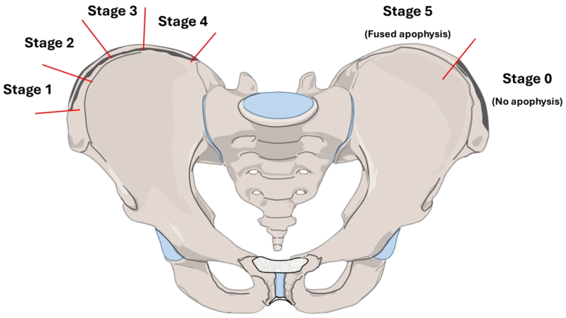
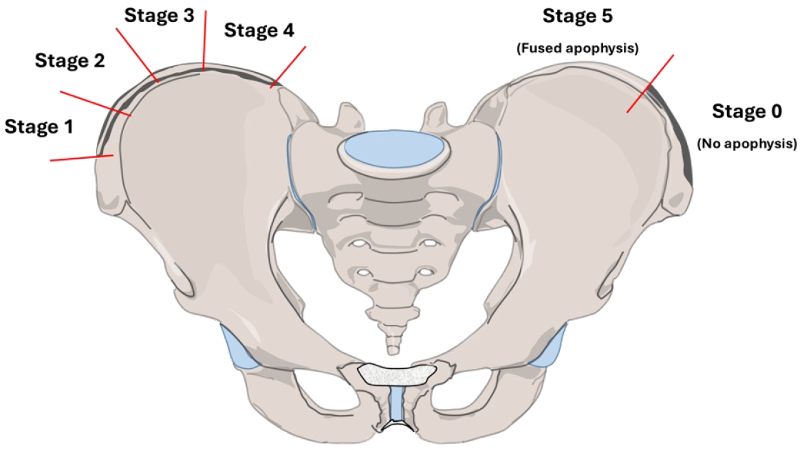
Radiographic indicators such as Risser status evaluate the ossification of the iliac crest apophysis on a scale of 0 to 5, to estimate skeletal maturity and remaining growth potential.56) The Risser classification is illustrated in Fig. 3.
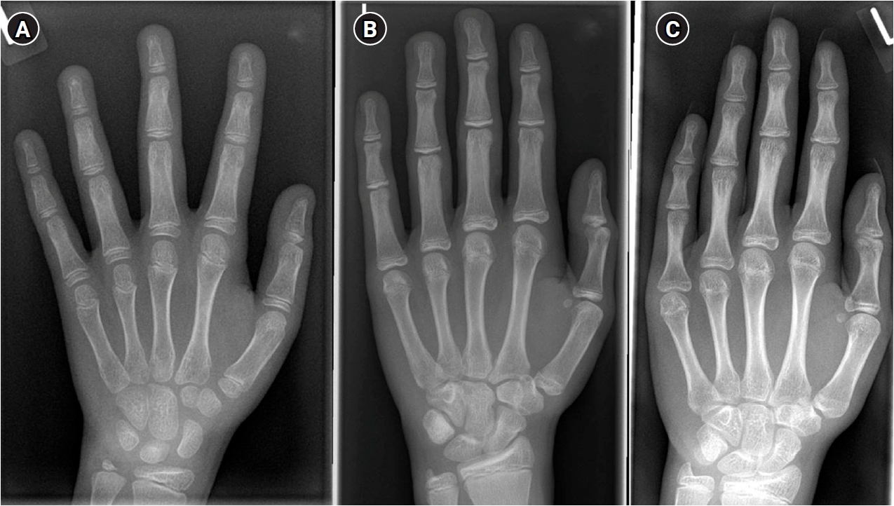
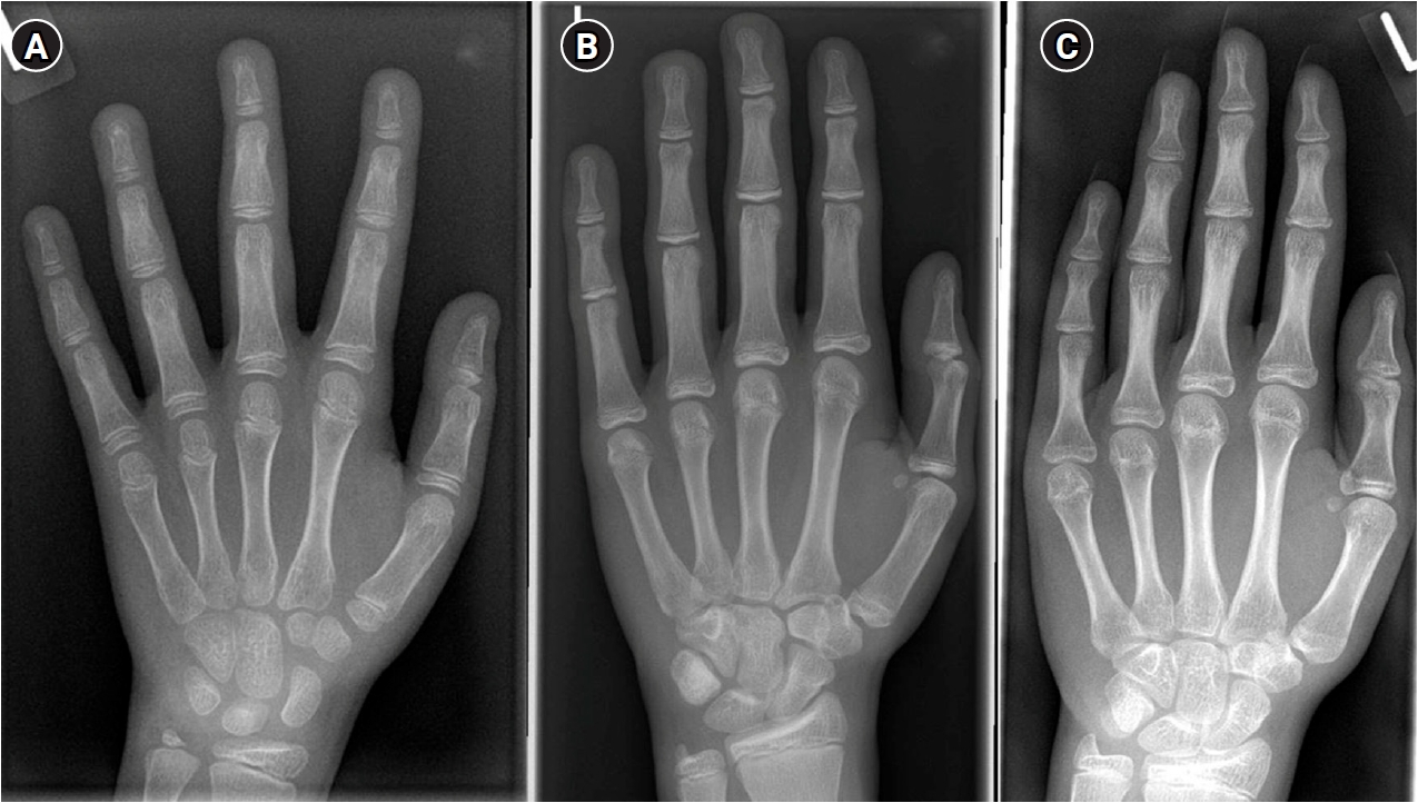
Additionally, the Sanders classification uses hand and wrist radiographs to provide a detailed assessment of skeletal age and is increasingly favored by surgeons to predict remaining growth potential, risk of progression and skeletal maturity with greater accuracy when compared to Risser staging.57-60) Representative radiographs of Sanders stages are shown in Fig. 4.
Management
Management of AIS is guided by the goals of preventing curve progression, correcting existing deformity, and minimizing long-term health impacts, while avoiding overtreatment in mild or non-progressive curves. Treatment depends largely on curve severity, remaining growth potential and evidence of progression. Multiple professional societies have published guidelines to assist clinical decision-making, including the Scoliosis Research Society (SRS), American Academy of Orthopaedic Surgeons (AAOS), and the International Society on Scoliosis Orthopaedic & Rehabilitation Treatment (SOSORT) for non-operative management. Treatment options include observation, bracing, and surgical intervention.
1. Conservative approaches in AIS
Observation with periodic monitoring is typically recommended for mild curves or patients nearing skeletal maturity with moderate curves. Specifically, adolescents with curves <20–25° who are still growing are initially observed.3,61) Curves up to ~30° may also be observed if skeletal maturity is near (Risser 4–5), as the risk of progression declines once growth plates fuse. Major guidelines, including those from the SRS and the AAOS, agree that bracing is not typically indicated for curves below 20–25° unless rapid progression occurs.62,63) The 2016 SOSORT guidelines and SRS criteria suggest bracing at 25° if significant growth remains; for instance, a 20° curve in a very immature child may warrant bracing if progression is noted.64)
Monitoring includes regular clinical assessment (including Adam’s forward bend test or scoliometer assessment) and standing radiographs at intervals, commonly every ~6 months during rapid growth.65,66) If a child is early in skeletal maturity (Risser 0–2) with a curve in the teens, follow-up occurs every ~6 months with X-rays to detect progression (≥5° increase). As growth slows, intervals lengthen to reduce radiation exposure.3,67) The typical cumulative radiation dose from surveillance has declined substantially with modern low-dose techniques, from historical levels of 10–20 mSv down to well under 5 mSv68,69), with patients treated with bracing receiving lower exposures than surgical patients70). Notwithstanding modern imaging techniques, breast cancer has been reported at 1.5–2.5 times the incidence in scoliosis patients subjected to multiple X-rays.70) Along with the Risser and Sander’s sign, skeletal maturity is assessed with menstrual status in girls, as growth deceleration occurs within 1–2 years of menarche.56)
2. Bracing
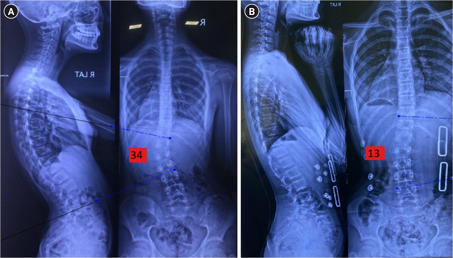
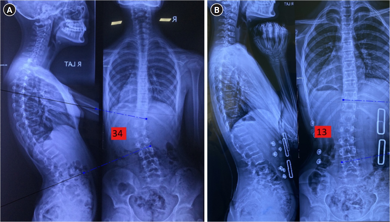
Bracing is the mainstay for AIS in growing patients with moderate curves.71) Classic SRS brace criteria include Cobb angles of ~25–40°, age ≥10, Risser 0–2, and, in girls, pre-menarche or within 1–2 years post-menarche.72) Bracing may be initiated earlier (20°) if rapid progression occurs (e.g., 15° to 22° within months in a Risser 0 patient). Conversely, curves >40–45° in immature patients may still be braced, though many ultimately require surgery. Bracing is ineffective in skeletally mature patients (Risser 5) or for severe curves (>50°), where surgical correction is more reliable. An example of out-of-brace and in-brace radiographs is shown in Fig. 5.
Several orthoses are available. The Boston brace, which is a type of thoracolumbosacral orthosis, is the most commonly used worldwide. It is a custom‑moulded plastic jacket worn for 18‒23 hours per day that applies padded pressure on convexities while leaving relief areas over concavities.73) Night time options such as the Charleston bending or Providence brace hyper-correct the curve only during sleep and are suited to flexible single lumbar or thoracolumbar patterns, giving better freedom during waking hours.74,75) Modern braces have a lower profile, can be worn under clothing, and increasingly incorporate embedded temperature or pressure sensors that record actual wear time and compliance.
Efficacy of bracing is supported by the landmark BrAIST randomized trial which demonstrated that roughly three‑quarters of braced patients avoided progression to the surgical threshold of 50°, compared with fewer than half of those simply observed, and patients who wore their brace for at least 13 hours daily achieved success rates exceeding 90%.71) Compliance is the key modifiable predictor yet also the greatest challenge; adherence often wanes during the teenage years.76) When wear is adequate and curves respond, bracing guides spinal growth, sparing many young people a fusion operation in the future.
Surgical Approaches
1. Indications for surgery
The primary indication for surgery in AIS is a curve that has reached a magnitude where progression is likely despite skeletal maturity, or a curve that already produces significant deformity or symptoms. In numeric terms, a Cobb angle of ≥45°–50° in a growing adolescent is a typical threshold for recommending spinal fusion surgery.77-79) This is because curves in this range have a high chance of exceeding 50°–60° by maturity and may continue to progress slowly in adulthood, risking future problems. Many surgeons use 50° as a clear cutoff at skeletal maturity since long-term studies show thoracic curves greater than 50° tend to progress in adult life at an average rate of approximately 1° per year.80) For large curves, fusion surgery may be the only way to achieve significant permanent correction.
2. Posterior spinal fusion
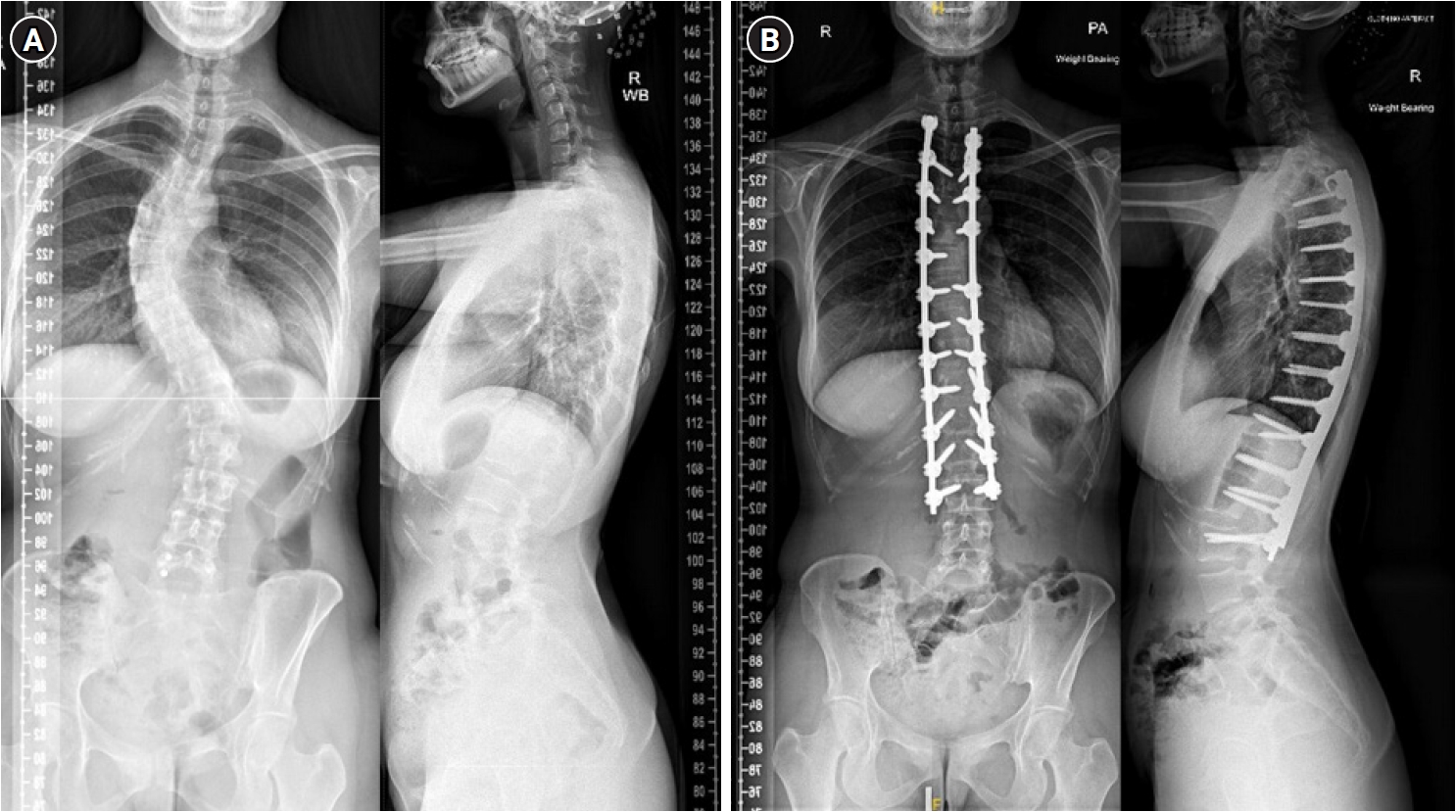
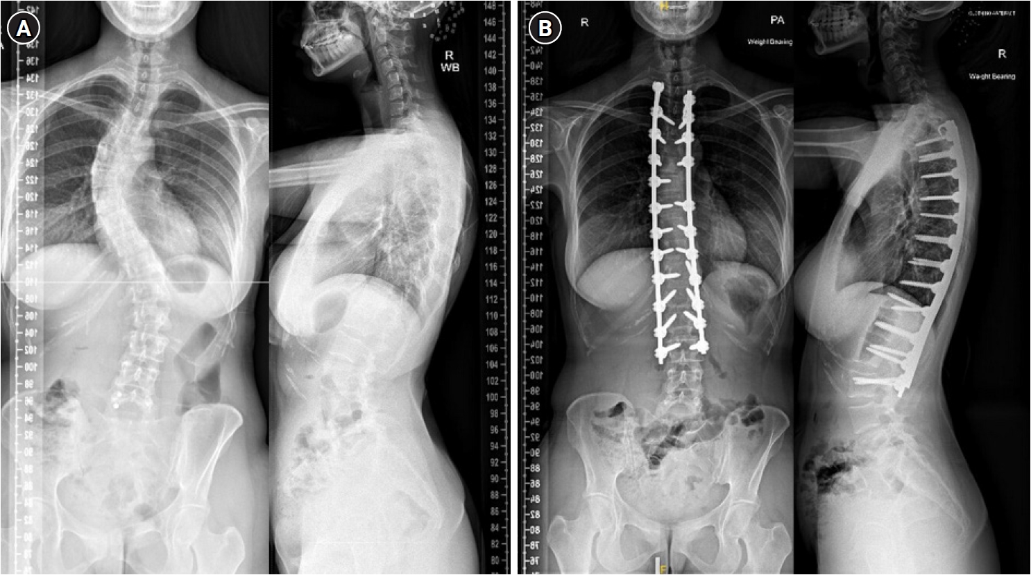
The posterior approach is the most popular surgical technique for AIS due to its effectiveness in achieving correction and stability. Through a midline incision along the back, the surgeon places metal implants, primarily pedicle screws, into the vertebrae spanning the curve. Two rigid rods, typically made of titanium or cobalt-chrome, are then connected to the implants and used to derotate and straighten the spine. Various corrective maneuvers, including rod derotation, direct vertebral rotation, translation, and compression/distraction techniques, are applied to achieve optimal alignment.81) Surgeons can then perform some fine tuning with “in-situ bending” of the rod. The operated vertebrae are then fused using bone grafts, which may be autografts harvested from local bone or allografts. Modern instrumentation allows for excellent curve correction, often reducing Cobb angle by 50%–80%, depending on flexibility.82) The goal is to preserve motion, by fusing the least amount of vertebral bodies while ensuring good correction and balance, and preventing subsequent progression of the non-fused segment, a decision guided by the curve type. In thoracic curves, selective thoracic fusion is a common strategy, in which only the primary thoracic curve is fused if the compensatory lumbar curve is flexible.83) The typical levels fused range from the upper end vertebra to the lower end vertebra of the structural curve, sometimes extending slightly beyond to maintain spinal balance. Representative preoperative and postoperative radiographs following posterior spinal fusion are shown in Fig. 6.
3. Anterior spinal fusion
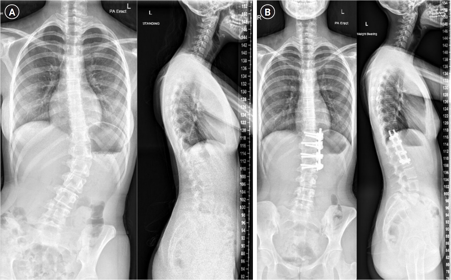
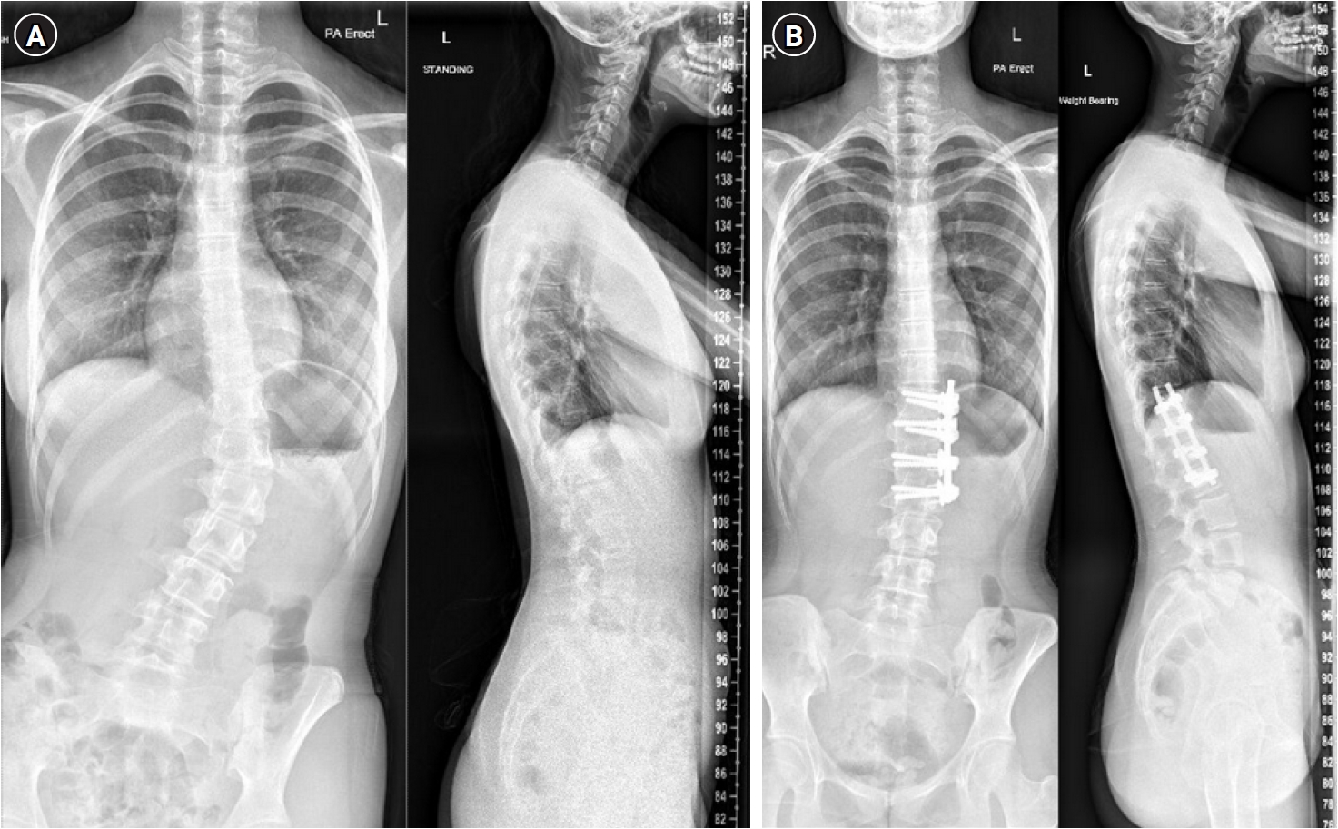
Historically, anterior spinal fusion was a more common approach before the widespread use of pedicle screw fixation, but it is now less frequently used for AIS.84,85) This technique involves an incision through the chest or flank to access the vertebral bodies. Screws are placed in the vertebral bodies, and a rod is connected to provide correction. In thoracolumbar curves, anterior fusion may allow the surgeon to fuse fewer levels while still achieving adequate correction.86) However, posterior pedicle screw fixation has largely replaced anterior approaches due to its ability to provide comparable or superior correction without the need for opening the chest. Representative preoperative and postoperative radiographs following anterior spinal fusion are shown in Fig. 7.
4. Vertebral body tethering
A newer surgical alternative for select cases is growth modulation without fusion, such as vertebral body tethering. This technique involves an anterior surgical approach where screws are placed in the vertebral bodies on the convex side of the curve and connected by a flexible polyethylene tether. Tension on the tether partially corrects the curve initially and subsequently modulates growth by slowing convex-side growth, allowing the concave side to catch up and straighten the spine over time. This method received U.S. Food and Drug Administration approval in 2019 for idiopathic scoliosis in skeletally immature patients (generally Risser 0–1) with curves ranging from 35° to 60°.87) While early results show promise in preserving spinal flexibility, tether breakage has been recognized as a common mechanical complication, long-term outcomes are still under investigation, and its use remains limited to specific indications.87,88)
Outcomes of Surgery
Surgical treatment of AIS is highly effective in achieving lasting correction and preventing further progression. Most patients report improved physical alignment and satisfaction with their appearance, including level shoulders and a reduced rib hump. A study on long-term quality of life after AIS surgery found significant postoperative improvements in pain, function, and self-image.89) By 12 months after fusion, most adolescents return to full activities, including sports, though restrictions may be advised for contact sports or extreme spinal motions based on individual recovery and surgeon recommendations.90)
Complications
Modern AIS surgery is safe, but as with any major surgery, there are risks. Neurological injury, caused by direct trauma, stretch, or ischemia is the most feared complication. However, it is extremely rare with current techniques and intraoperative neuromonitoring, occurring in approximately 0.2% of spinal deformity surgeries.91) Major blood loss is rare, mitigated through surgical technique, cell-saver technology and antifibrinolytic agents such as tranexamic acid.92,93) Infection rates range from 1% to 5%, with deep wound infection necessitating extended antibiotic therapy and implant removal.94) Instrumentation-related issues, such as rod breakage or screw loosening, have low incidence due to improved implant designs.95,96) Pseudarthrosis is rare in adolescents (~1% or less) due but may require revision surgery if it leads to pain or rod breakage.97) Degenerative disc disease in the unfused spinal column distal to fused segments is a further complication, with these rates increasing in years following the surgery; the rates remain higher in non-operative patients but are not absent with surgical intervention.6,98) Patients with fusion levels at L3 or above exhibit less advanced disc degeneration over time which likely translates into lower long term back pain rates.99) While scarring is an unavoidable consequence, posterior scars typically heal well and can be concealed by clothing.
Conclusion
AIS affects 2%–4% of adolescents, with females disproportionately experiencing curve progression.3) It is a multifactorial disorder driven by polygenic, hormonal, and biomechanical influences.100) Early detection is crucial, as timely bracing during growth can effectively alter the natural history of moderate curves, while modern surgical techniques safely correct severe deformities. Despite these advances, challenges remain in fully understanding AIS etiology and predicting progression. Future interdisciplinary strategies, including personalized risk profiling, novel non-fusion treatments, and enhanced screening technologies, will be key to ensuring that each patient receives the optimal care for a healthy, active life.
NOTES
Author contributions
Conceptualization: CHL, HIB, CTB, MJ. Data curation: CHL, HIB, CTB, MJ. Formal analysis: CHL, HIB. Investigation: CHL, HIB, CTB, MM, MK, PD, SCS, PJ, MJ. Methodology: CHL, HIB, CTB, MJ. Project administration: CHL, HIB, CTB. Resources: CHL, HIB. Supervision: CHL, HIB, CTB, JM, GM, MJ. Validation: CHL, HIB, CTB, SH, JM, GM, MJ. Visualization: CHL, HIB, CTB, PJ. Writing - original draft: CHL, HIB, CTB, MM, MK, PD, SCS, PJ. Writing - review & editing: CHL, HIB, CTB, MM, MK, HT, PD, SCS, PJ, SH, DSM, JM, GM, MJ.
Conflict of interest
The authors have no conflicts of interest to declare.
Funding
None.
Acknowledgments
None.
Fig. 1.
Typical standing back image (A) and forward bending back image (B) of a 15-year-old patient with a thoracolumbar curve.
Fig. 2.
An anterior posterior, lateral, and posteroanterior radiograph including the cervical spine and both iliac crests in a 14-year-old female patient with a right thoracic curve.
Fig. 3.
The Risser classification. Stage 0: no ossification of the apophysis, Stage I: apophysis over 25% of the iliac crest, II: apophysis over 25%–50% of the iliac crest, III: apophysis over 50%–75% of the iliac crest, IV: apophysis over >75% of the iliac crest, V: complete ossification and fusion of the iliac crest apophysis.
Fig. 4.
Radiographs showing different Sanders stages. (A) Sanders 2. (B) Sanders 3. (C) Sanders 5. Adapted from Braun et al. Life (Basel) 2023;13:1341,60) according to Creative Commons license.
Fig. 5.
Out-of-brace X-ray (A) compared to an In-brace X-ray (B) with Cobb angle measurements highlighted in red in a 13-year-old female patient with a type 5 curve.
Fig. 6.
Anteroposterior and lateral X-rays pre-posterior fusion (A), and anteroposterior and lateral X-rays post-posterior fusion (B) of a 17-year-old male patient with a double thoracic curve.
Fig. 7.
Anteroposterior and lateral X-rays pre-anterior fusion (A), and anteroposterior and lateral X-rays post-anterior fusion (B) of a 15-year-old female patient with a left type 5 curve.
Table 1.
Summary of adolescent idiopathic scoliosis epidemiology
1. Aulia TN, Djufri D, Gatam L, Yaman A. Etiopathogenesis of adolescent idiopathic scoliosis (AIS): role of genetic and environmental factors. Narra J 2023;3:e217.
5. Mitsiaki I, Thirios A, Panagouli E, et al. Adolescent idiopathic scoliosis and mental health disorders: a narrative review of the literature. Children (Basel) 2022;9:597.
7. Plaszewski M, Grantham W, Jespersen E. Screening for scoliosis: new recommendations, old dilemmas, no straight solutions. World J Orthop 2020;11:364-79.
8. Weiss HR, Karavidas N, Moramarco M, Moramarco K. Long-term effects of untreated adolescent idiopathic scoliosis: a review of the literature. Asian Spine J 2016;10:1163-9.
9. Weinstein SL, Dolan LA, Spratt KF, Peterson KK, Spoonamore MJ, Ponseti IV. Health and function of patients with untreated idiopathic scoliosis: a 50-year natural history study. JAMA 2003;289:559-67.
11. Wang L, Wang YP, Yu B, et al. Relation between self-image score of SRS-22 with deformity measures in female adolescent idiopathic scoliosis patients. Orthop Traumatol Surg Res 2014;100:797-801.
13. Luk KD, Lee CF, Cheung KM, et al. Clinical effectiveness of school screening for adolescent idiopathic scoliosis: a large population-based retrospective cohort study. Spine (Phila Pa 1976) 2010;35:1607-14.
14. Komang-Agung IS, Dwi-Purnomo SB, Susilowati A. Prevalence rate of adolescent idiopathic scoliosis: results of school-based screening in Surabaya, Indonesia. Malays Orthop J 2017;11:17-22.
15. Daruwalla JS, Balasubramaniam P, Chay SO, Rajan U, Lee HP. Idiopathic scoliosis. Prevalence and ethnic distribution in Singapore schoolchildren. J Bone Joint Surg Br 1985;67:182-4.
16. Suh SW, Modi HN, Yang JH, Hong JY. Idiopathic scoliosis in Korean schoolchildren: a prospective screening study of over 1 million children. Eur Spine J 2011;20:1087-94.
17. Grauers A, Danielsson A, Karlsson M, Ohlin A, Gerdhem P. Family history and its association to curve size and treatment in 1,463 patients with idiopathic scoliosis. Eur Spine J 2013;22:2421-6.
19. Grivas TB, Vasiliadis E, Mouzakis V, Mihas C, Koufopoulos G. Association between Adolescent Idiopathic Scoliosis prevalence and age at menarche in different geographic latitudes. Scoliosis 2006;1:9.
21. Tang NL, Yeung HY, Hung VW, et al. Genetic epidemiology and heritability of AIS: a study of 415 Chinese female patients. J Orthop Res 2012;30:1464-9.
22. Peng Y, Wang SR, Qiu GX, Zhang JG, Zhuang QY. Research progress on the etiology and pathogenesis of adolescent idiopathic scoliosis. Chin Med J (Engl) 2020;133:483-93.
25. Perez-Machado G, Berenguer-Pascual E, Bovea-Marco M, et al. From genetics to epigenetics to unravel the etiology of adolescent idiopathic scoliosis. Bone 2020;140:115563.
26. Ward K, Ogilvie J, Argyle V, et al. Polygenic inheritance of adolescent idiopathic scoliosis: a study of extended families in Utah. Am J Med Genet A 2010;152A:1178-88.
27. Londono D, Kou I, Johnson TA, et al. A meta-analysis identifies adolescent idiopathic scoliosis association with LBX1 locus in multiple ethnic groups. J Med Genet 2014;51:401-6.
28. Ushiki A, Sheng RR, Zhang Y, et al. Deletion of Pax1 scoliosis-associated regulatory elements leads to a female-biased tail abnormality. Cell Rep 2024;43:113907.
29. Xu L, Dai Z, Xia C, et al. Asymmetric expression of Wnt/B-catenin pathway in AIS: primary or secondary to the curve? Spine (Phila Pa 1976) 2020;45:E677-83.
30. Singh H, Shipra , Gupta M, et al. SOX9 gene shows association with adolescent idiopathic scoliosis predisposition in northwest Indians. Eur J Med Res 2024;29:66.
34. Borysiak K, Janusz P, Andrusiewicz M, et al. CHD7 gene polymorphisms in female patients with idiopathic scoliosis. BMC Musculoskelet Disord 2020;21:18.
36. Li L, Wong MS. The application of machine learning methods for predicting the progression of adolescent idiopathic scoliosis: a systematic review. Biomed Eng Online 2024;23:80.
37. McCallum-Loudeac J, Wilson MJ. Adolescence and scoliosis: deciphering the complex biology of puberty and scoliosis. In: Kusumi K, Dunwoodie SL, editors. The genetics and development of scoliosis. Springer International Publishing; 2018. p. 179-93.
38. Petrosyan E, Fares J, Ahuja CS, et al. Genetics and pathogenesis of scoliosis. N Am Spine Soc J 2024;20:100556.
43. Gardner A, Berryman F, Pynsent P. The kyphosis–lordosis difference parameter and its utility in understanding the pathogenesis of adolescent idiopathic scoliosis. BMC Res Notes 2022;15:178.
44. Schlosser TP, van Stralen M, Brink RC, et al. Three-dimensional characterization of torsion and asymmetry of the intervertebral discs versus vertebral bodies in adolescent idiopathic scoliosis. Spine (Phila Pa 1976) 2014;39:E1159-66.
45. Brink RC, Schlosser TPC, van Stralen M, et al. Anterior-posterior length discrepancy of the spinal column in adolescent idiopathic scoliosis-a 3D CT study. Spine J 2018;18:2259-65.
48. Tanaka A. A histopathological study on the intervertebral discs of idiopathic and paralytic scoliosis: abnormalities in transition from the notochordal nucleus to the fibrocartilaginous nucleus. Nihon Seikeigeka Gakkai Zasshi 1986;60:1227-38.
49. Yeung KH, Man GC, Deng M, et al. Morphological changes of intervertebral disc detectable by T2-weighted MRI and its correlation with curve severity in adolescent idiopathic scoliosis. BMC Musculoskelet Disord 2022;23:655.
50. Yu SH, Ng CM, Cheung JP, Shea GK. Post-maturity progression in adolescent idiopathic scoliosis curves of 40° to 50°. J Bone Joint Surg Am 2023;105:277-85.
51. Llopis-Ibor CI, Mariscal G, de la Rubia Orti JE, Barrios C. Incidence of vitamin D deficiency in adolescent idiopathic scoliosis: a meta-analysis. Front Endocrinol (Lausanne) 2023;14:1250118.
52. Sarioglu O, Gezer S, Sarioglu FC, et al. Evaluation of vertebral bone mineral density in scoliosis by using quantitative computed tomography. Pol J Radiol 2019;84:e131-5.
53. Sun D, Ding Z, Hai Y, Cheng Y. Advances in epigenetic research of adolescent idiopathic scoliosis and congenital scoliosis. Front Genet 2023;14:1211376.
55. Lenke LG, Betz RR, Harms J, et al. Adolescent idiopathic scoliosis: a new classification to determine extent of spinal arthrodesis. J Bone Joint Surg Am 2001;83:1169-81.
56. Hacquebord JH, Leopold SS. In brief: the Risser classification: a classic tool for the clinician treating adolescent idiopathic scoliosis. Clin Orthop Relat Res 2012;470:2335-8.
57. Minkara A, Bainton N, Tanaka M, et al. High risk of mismatch between Sanders and Risser staging in adolescent idiopathic scoliosis: are we guiding treatment using the wrong classification? J Pediatr Orthop 2020;40:60-4.
58. Cheung PWH, Cheung JPY. Does the use of Sanders staging and distal radius and ulna classification avoid mismatches in growth assessment with Risser staging alone? Clin Orthop Relat Res 2021;479:2516-30.
59. Vira S, Husain Q, Jalai C, et al. The interobserver and intraobserver reliability of the Sanders classification versus the Risser stage. J Pediatr Orthop 2017;37:e246-9.
60. Braun S, Brenneis M, Schonnagel L, Caffard T, Diaremes P. Surgical treatment of spinal deformities in pediatric orthopedic patients. Life (Basel) 2023;13:1341.
61. Dunn J, Henrikson NB, Morrison CC, Nguyen M, Blasi PR, Lin JS. Screening for adolescent idiopathic scoliosis: a systematic evidence review for the U.S. Preventive Services Task Force. Agency for Healthcare Research and Quality (US); 2018.
62. Karavidas N. Bracing in the treatment of adolescent idiopathic scoliosis: evidence to date. Adolesc Health Med Ther 2019;10:153-72.
64. Negrini S, Hresko TM, O'Brien JP, Price N, SOSORT Boards; SRS Non-Operative Committee. Recommendations for research studies on treatment of idiopathic scoliosis: consensus 2014 between SOSORT and SRS Non-Operative Management Committee. Scoliosis 2015;10:8.
65. Cote P, Kreitz BG, Cassidy JD, Dzus AK, Martel J. A study of the diagnostic accuracy and reliability of the Scoliometer and Adam’s forward bend test. Spine (Phila Pa 1976) 1998;23:796-802.
66. Peeters CM, van Hasselt AJ, Wapstra FH, Jutte PC, Kempen DH, Faber C. In-brace versus out-of-brace protocol for radiographic follow-up of patients with idiopathic scoliosis: a retrospective study. Children (Basel) 2022;9:465.
69. Cool J, Streekstra GJ, van Schuppen J, Stadhouder A, van den Noort JC, van Royen BJ. Estimated cumulative radiation exposure in patients treated for adolescent idiopathic scoliosis. Eur Spine J 2023;32:1777-86.
70. Doody MM, Lonstein JE, Stovall M, Hacker DG, Luckyanov N, Land CE. Breast cancer mortality after diagnostic radiography: findings from the U.S. scoliosis cohort study. Spine (Phila Pa 1976) 2000;25:2052-63.
74. Gepstein R, Leitner Y, Zohar E, et al. Effectiveness of the Charleston bending brace in the treatment of single-curve idiopathic scoliosis. J Pediatr Orthop 2002;22:84-7.
75. Yrjonen T, Ylikoski M, Schlenzka D, Kinnunen R, Poussa M. Effectiveness of the providence nighttime bracing in adolescent idiopathic scoliosis: a comparative study of 36 female patients. Eur Spine J 2006;15:1139-43.
76. Takemitsu M, Bowen JR, Rahman T, Glutting JJ, Scott CB. Compliance monitoring of brace treatment for patients with idiopathic scoliosis. Spine (Phila Pa 1976) 2004;29:2070-4.
80. Danielsson AJ. Natural history of adolescent idiopathic scoliosis: a tool for guidance in decision of surgery of curves above 50°. J Child Orthop 2013;7:37-41.
82. Mirzashahi B, Moosavi M, Rostami M. Outcome of posterior-only approach for severe rigid scoliosis: a retrospective report. Int J Spine Surg 2020;14:232-8.
86. Hirase T, Ling JF, Haghshenas V, et al. Anterior versus posterior spinal fusion for Lenke type 5 adolescent idiopathic scoliosis: a systematic review and meta-analysis of comparative studies. Spine Deform 2022;10:267-81.
87. Roser MJ, Askin GN, Labrom RD, Zahir SF, Izatt M, Little JP. Vertebral body tethering for idiopathic scoliosis: a systematic review and meta-analysis. Spine Deform 2023;11:1297-307.
88. Baroncini A, Trobisch P, Eschweiler J, Migliorini F. Analysis of the risk factors for early tether breakage following vertebral body tethering in adolescent idiopathic scoliosis. Eur Spine J 2022;31:2348-54.
89. Mens RH, Bisseling P, de Kleuver M, van Hooff ML. Relevant impact of surgery on quality of life for adolescent idiopathic scoliosis: a registry-based two-year follow-up cohort study. Bone Joint J 2022;104B:265-73.
90. Barile F, Ruffilli A, Manzetti M, et al. Resumption of sport after spinal fusion for adolescent idiopathic scoliosis: a review of the current literature. Spine Deform 2021;9:1247-51.
91. Leong JJ, Curtis M, Carter E, Cowan J, Lehovsky J. Risk of neurological injuries in spinal deformity surgery. Spine (Phila Pa 1976) 2016;41:1022-7.
92. Stone N, Sardana V, Missiuna P. Indications and outcomes of Cell Saver in adolescent scoliosis correction surgery: a systematic review. Spine (Phila Pa 1976) 2017;42:E363-70.
93. Shrestha IK, Ruan TY, Lin L, et al. The efficacy and safety of high-dose tranexamic acid in adolescent idiopathic scoliosis: a meta-analysis. J Orthop Surg Res 2021;16:53.
94. Aleissa S, Parsons D, Grant J, Harder J, Howard J. Deep wound infection following pediatric scoliosis surgery: incidence and analysis of risk factors. Can J Surg 2011;54:263-9.
95. Uehara M, Takahashi J, Ikegami S, et al. Pedicle screw loosening after posterior spinal fusion for adolescent idiopathic scoliosis in upper and lower instrumented vertebrae having major perforation. Spine (Phila Pa 1976) 2017;42:1895-900.
96. Ohrt-Nissen S, Dahl B, Gehrchen M. Choice of rods in surgical treatment of adolescent idiopathic scoliosis: What are the clinical implications of biomechanical properties?: a review of the literature. Neurospine 2018;15:123-30.
98. Lonner BS, Ren Y, Upasani VV, et al. Disc degeneration in unfused caudal motion segments ten years following surgery for adolescent idiopathic scoliosis. Spine Deform 2018;6:684-90.
99. Akazawa T, Kotani T, Sakuma T, et al. Spinal fusion on adolescent idiopathic scoliosis patients with the level of L4 or lower can increase lumbar disc degeneration with sagittal imbalance 35 years after surgery. Spine Surg Relat Res 2017;1:72-7.
Adolescent Idiopathic Scoliosis: A Comprehensive Review of Epidemiology, Etiology, and Interventions
Fig. 1. Typical standing back image (A) and forward bending back image (B) of a 15-year-old patient with a thoracolumbar curve.
Fig. 2. An anterior posterior, lateral, and posteroanterior radiograph including the cervical spine and both iliac crests in a 14-year-old female patient with a right thoracic curve.
Fig. 3. The Risser classification. Stage 0: no ossification of the apophysis, Stage I: apophysis over 25% of the iliac crest, II: apophysis over 25%–50% of the iliac crest, III: apophysis over 50%–75% of the iliac crest, IV: apophysis over >75% of the iliac crest, V: complete ossification and fusion of the iliac crest apophysis.
Fig. 4. Radiographs showing different Sanders stages. (A) Sanders 2. (B) Sanders 3. (C) Sanders 5. Adapted from Braun et al. Life (Basel) 2023;13:1341,60) according to Creative Commons license.
Fig. 5. Out-of-brace X-ray (A) compared to an In-brace X-ray (B) with Cobb angle measurements highlighted in red in a 13-year-old female patient with a type 5 curve.
Fig. 6. Anteroposterior and lateral X-rays pre-posterior fusion (A), and anteroposterior and lateral X-rays post-posterior fusion (B) of a 17-year-old male patient with a double thoracic curve.
Fig. 7. Anteroposterior and lateral X-rays pre-anterior fusion (A), and anteroposterior and lateral X-rays post-anterior fusion (B) of a 15-year-old female patient with a left type 5 curve.
Fig. 1.
Fig. 2.
Fig. 3.
Fig. 4.
Fig. 5.
Fig. 6.
Fig. 7.
Adolescent Idiopathic Scoliosis: A Comprehensive Review of Epidemiology, Etiology, and Interventions


 , Halil Ibrahim Bulut, M.D.2)