Purpose This retrospective study investigated the distinct clinical and radiographic drivers of early- versus late-onset proximal junctional kyphosis (PJK) following multilevel thoracolumbar (TL) fusion.
Methods After applying the exclusion criteria (spinal infection, neuromuscular disease, age <50 years), the analysis included 136 patients who underwent ≥4-level TL fusion and were followed up for a minimum of 2 years. PJK was classified as early (≤6 months) or late (>6 months) onset. Patient-related factors, surgical variables, sagittal spinopelvic parameters, and preoperative magnetic resonance imaging findings were analyzed using multivariate logistic regression to identify independent predictors of early PJK.
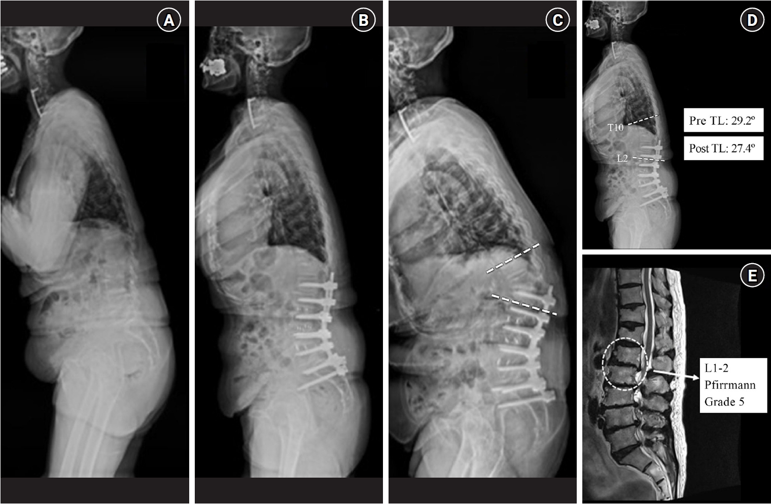
Results Among 24 patients (17.6%) who developed PJK, the early and late-onset groups included 13 and 11 patients, respectively. The early PJK group exhibited significantly greater preoperative and postoperative TL angles compared with the late group (preoperative: 23.03±13.83° vs. 9.67±9.67°, p=0.024; postoperative: 19.6±6.95° vs. 6.95±6.35°, p<0.001). The Pfirrmann grade of the L1–2 intervertebral disc was significantly higher in the early PJK group (3.92±0.95 vs. 2.81±0.60, p=0.006). No surgical variables differed significantly between the groups. Multivariate analysis confirmed greater postoperative TL angle and more advanced L1–2 disc degeneration as independent predictors of early PJK.
Conclusion Early-onset PJK following multilevel TL fusion is primarily driven by regional biomechanical vulnerabilities, specifically residual postoperative TL kyphosis and advanced adjacent L1–2 disc degeneration, rather than by surgical variables. Meticulous evaluation of regional TL alignment and adjacent disc health during surgical planning is critical for risk stratification and prevention of early junctional failure.
Chronic low back pain is a common cause of disability causing major socioeconomic consequences. Recent advances in disc biology and tissue engineering techniques enable a new emerging field of biologic treatments for degenerative disc disease. These new treatment modalities aim to achieve structural and functional restoration of the degenerated discs by introducing protein, cells, genetic modifications of resident disc cells or exogenous cultured cells, and use of biomaterials. So far, these techniques have been successfully applied to treat degenerated discs in preclinical setting, including in vitro or in animal studies. Application of these treatment modalities for degenerative disc disease should be individualized according to the degree of disc degeneration. For the successful application in clinical field, the biologic treatment should achieve the functional restoration of the disc, resulting in pain regulation.
Introduction Degenerative change of the vertebral disc is known to be caused by inherited factors, aging and mechanical and environmental factors and it progresses due to the decreasing amount of proteoglycan in the vertebral disc. Resolving the imbalance between the synthesis and break down of proteoglycan is an important strategy for slowing additional degeneration and increasing regeneration of the disc.
Body: To regenerate vertebral discs, many attempt have been made to get curative value such as direct injection of growth factors like BMP-2, BMP-7, IGF-1, TGF-β1, GDF-5 and FGF into the disc or transplantation of cytokine for improving the composition of proteoglycan. But these proteins have a short acting time and so repeated transplantation is necessary even if transplantation into the disc is done. So, much research has been done on genetic transplantation, which may improve certain proteins. Other trials of transplantation of nucleus pulposus cells, combinations of stem cells with scaffold and amalgamation with genetic care have also been done. But the safety of these therapies has not yet been completely established.
Conclusion Biological therapy for degenerative disc disease has many benefits compared with the conventional therapies. Hereafter, according to additional research, the clinical indications for biological therapies are expected to increase. Clinical applications should be performed after securing enough results for establishing the safety and effectiveness of these new procedures.


 First
First Prev
Prev